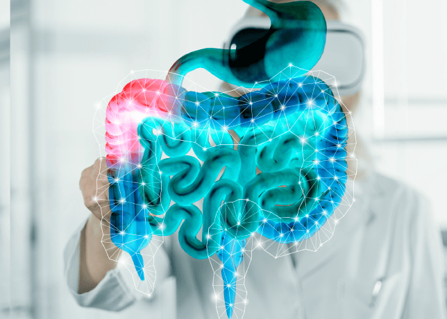
Enhancing Colon Polyp Detection through Deep Learning: A YOLO-Based Approach
- [printfriendly]
Colorectal Cancer (CRC) is characterized by its notable status as the third most lethal form of cancer. According to the cited source, colon cancer demonstrates a five-year survival rate of roughly 68%, but stomach cancer exhibits a comparatively lower survival rate of 44% [1]. In the pursuit of reducing mortality rates related to colorectal cancer (CRC), an approach of significant efficacy involves the detection and elimination of precancerous abnormalities.
The identification of colon polyps at an early stage is crucial due to their capacity to develop into advanced colorectal cancer (CRC). Hence, the early identification of CRC is of utmost importance for preserving lives.
Besides implementing lifestyle modifications, regular colon screening plays a crucial role in the prevention of colorectal cancer (CRC). Numerous empirical studies have yielded compelling findings that provide support to the proposition that the adoption of population screening holds promise for improving prognosis and potentially reducing the prevalence of colorectal cancer [2]. The colonoscopy procedure entails the inspection and intervention of the colon via a flexible endoscope, administered by a certified endoscopist. The procedure is regarded as an intrusive medical intervention. The diagnostic tool utilized for the prompt detection and subsequent removal of polyps during colon examination is usually acknowledged as the foremost approach[3].
As a result, gastroenterologists commonly choose colonoscopic screening as the preferred approach. Polyps are defined as anomalous formations of tissue that extend outward from the mucosal membrane and can manifest at any site within the gastrointestinal (GI) tract. Nevertheless, these lesions are primarily identified in the colorectal region and are well acknowledged as precursors of colorectal cancer (CRC) [4], [5]. There exist two main classifications of polyps: pedunculated, characterized by a well-defined stalk, and sessile, lacking a discernible stalk. Colorectal polyps can be classified into two primary categories: nonneoplastic and neoplastic. Non-neoplastic polyps can be categorized into three distinct groups: hyperplastic, inflammatory, and hamartomatous polyps. It is crucial to acknowledge that these polyps are of a benign nature and do not possess any inherent risk of progressing into malignant tumors[6]. Neoplastic polyps can be categorized into two primary classifications: adenomas and serrated polyps. It is noteworthy to acknowledge that both categories possess the potential to develop into malignant tumors. Colorectal polyps can be classified into three discrete categories according to their dimensions: diminutive (≤5mm), small (6 to 9 mm), and advanced (large) (≥10mm). The user's text does not contain any information to rewrite in an academic manner.
In general, the identification and removal of bigger polyps is usually straightforward; nevertheless, it is important to note that there is a significant risk associated with small and minuscule colorectal polyps [7]. Polypectomy is a procedure employed to eliminate small and minuscule polyps. There are five different procedures used to remove small polyps, including cold forceps polypectomy, hot forceps polypectomy, cold snare polypectomy, hot snare polypectomy, and endoscopic mucosal resection [8]. Among the several approaches considered, cold snare polypectomy emerges as the optimal selection for the excision of tiny colorectal polyps [9]. The colonoscopy process, because to its invasive nature, requires careful intestinal preparation and the utilization of air insufflation throughout the inspection [10].
The aforementioned technique is characterized by both a significant financial investment and a substantial time requirement. However, it is worth noting that around 20% of polyps remain undetected during medical examinations. The risk of developing cancer is dependent on the skill and expertise of the endoscopist in detecting polyps [11]. Recent studies have provided evidence that the utilization of innovative endoscopic instruments and diagnostic techniques has resulted in improvements in both adenoma detection rates and polyp detection rates [12], [13]. However, the matter of disregarded polyps continues to be a concern.
Colonoscopy recordings are routinely documented by clinical centers, resulting in the accumulation of a significant amount of colonoscopy data. Unfortunately, the potential of this important data remains untapped due to the labor-intensive nature of its analysis by endoscopists [14]. As a result, the importance of secondary video checks is often overlooked, which might potentially result in the failure to identify early-stage irregularities. The incorporation of automated data curation and video data annotation is crucial in the development of reliable Computer-Aided Diagnosis (CADx) systems, which have the potential to greatly improve the thorough assessment of clinical endoscopy [15].
A subset of the collected colonoscopy data can be meticulously analyzed in order to develop computer-assisted systems that possess the capability to autonomously identify and outline polyps. This might be accomplished either during the clinical procedure or in the subsequent step of reporting. In order to ensure the robustness of a system, it is important to integrate data variances related to patients, the specificities of the endoscopic operation, and variations stemming from diverse endoscope manufacturers[16].
In spite of the recent developments in computer vision and system design, which have facilitated the development of accurate and efficient systems, it is crucial to acknowledge that these progressions are heavily contingent upon the availability of data. A considerable amount of data is necessary for the implementation of numerous contemporary methodologies. One notable obstacle in accelerating algorithm development in this field is the limited availability of publicly accessible datasets [17]. The curation of medical datasets is typically a multifaceted undertaking that requires specialized knowledge in the respective discipline.
Obtaining a consensus among multiple experts regarding the ground truth labels for a given dataset poses still another challenge. In the context of colonoscopy, it is common for tiny polyps or flat/sessile polyps to go unnoticed throughout the process[18], making their observation challenging even with manual labeling. Additional obstacles arise from the inherent heterogeneity across patients and the existence of polyps with diverse characteristics, such as varying sizes, forms, textures, colors, and orientations. Hence, it is imperative to address the several issues that commonly arise during routine colonoscopy when engaging in polyp data curation and the development of automated systems for this procedure. Deep Learning (DL)-based algorithms for automatic polyp recognition and segmentation have significant efficacy in both colonoscopy images and colonoscopy films, as demonstrated by previous studies [19], [20]. Ideally, the automatic computer-aided diagnosis (CADx) systems designed for the detection, localization, and segmentation of polyps should possess the following characteristics: 1) Consistent performance and enhanced robustness to account for patient variability, ensuring the system consistently generates reliable outputs. 2) High overall performance that exceeds the established benchmarks for algorithms, indicating superior diagnostic accuracy. 3) Real-time performance capabilities that are essential for practical implementation in clinical settings, allowing for timely and efficient diagnosis. 4) An intuitive and user-friendly interface that provides clinically interpretable outputs, facilitating ease of use for medical professionals. Scaling this up to a cohort of population size is a highly resource-intensive process that involves significant costs. In order to initiate the process, our focus is directed on the identification, positioning, and division of colorectal polyps, which are recognized as precursors of colorectal cancer (CRC). The rationale behind commencing with this particular scenario is from the fact that a significant proportion of colon malignancies, approximately 20%, originate from benign adenomatous polyps that have dysplastic cellular characteristics. The prevention of cancer through the detection and removal of polyps is crucial, as the likelihood of developing colorectal cancer (CRC) within the subsequent 60 months after a colonoscopy is primarily contingent upon the proficiency of the endoscopist in identifying polyps[21]. The identification and localization of polyps are typically crucial in routine surveillance and for assessing the polyp burden of patients at the conclusion of surveillance. Additionally, pixel-wise segmentation is essential for automating the delineation of polyp boundaries during surgical procedures or radio-frequency ablations. This work presents an evaluation of deep learning (DL) approaches for both detection and segmentation tasks on the Kvasir-SEG dataset. The detection task involves localizing objects using bounding box detection, while the segmentation task involves pixel-wise classification or semantic segmentation. The purpose of this evaluation is to establish a comprehensive standard for colonoscopy images[22]. The primary objective of this study is to build a robust benchmark that can be used to compare and evaluate the performance of various computer vision algorithms, in order to identify the most successful ones. The contributions made by our team can be succinctly summarized as follows:
- We conducted training sessions using various models, including YOLOv5 and YOLOv8 (the most recent iterations of the You Only Look/Live Once model), on the Kvasir-SEG dataset to identify the most accurate model.
- A thorough evaluation of state-of-the-art computer vision baseline methods on the Kvasir-SEG dataset is provided. The top-performing approaches demonstrate real-time capabilities in both polyp detection and localization.
- We assessed the detection and localization performance using established computer vision metrics.
- We offer an in-depth analysis, with particular emphasis on both the most successful and the least effective scenarios. This approach allows us to scrutinize the factors contributing to method success and those responsible for shortcomings, ultimately expediting algorithm development
Reference:
[1] J. Asplund, J. H. Kauppila, F. Mattsson, and J. Lagergren, “Survival trends in gastric adenocarcinoma: a population-based study in Sweden,” Ann Surg Oncol, vol. 25, pp. 2693–2702, 2018.
[2] Ø. Holme, M. Bretthauer, A. Fretheim, J. Odgaard‐Jensen, and G. Hoff, “Flexible sigmoidoscopy versus faecal occult blood testing for colorectal cancer screening in asymptomatic individuals,” Cochrane Database of Systematic Reviews, no. 9, 2013.
[3] M. A. Deif, M. A. A. Eldosoky, H. W. Gomma, A. M. El-Garhy, and A. S. Ell-Azab, “Adaptive Neuro-Fuzzy Inference System Controller Technique for Lower Urinary Tract System Disorders,” J Clin Eng, vol. 40, no. 3, pp. 135–143, 2015.
[4] D. Jha et al., “Resunet++: An advanced architecture for medical image segmentation,” in 2019 IEEE international symposium on multimedia (ISM), IEEE, 2019, pp. 225–2255.
[5] R. G. Holzheimer and J. A. Mannick, “Surgical treatment: evidence-based and problem-oriented,” 2001.
[6] M. A. Deif, H. Attar, A. Amer, I. A. Elhaty, M. R. Khosravi, and A. A. A. Solyman, “Diagnosis of Oral Squamous Cell Carcinoma Using Deep Neural Networks and Binary Particle Swarm Optimization on Histopathological Images: An AIoMT Approach,” 2022.
[7] J. Lee, “Resection of diminutive and small colorectal polyps: What is the optimal technique?,” Clin Endosc, vol. 49, no. 4, pp. 355–358, 2016.
[8] P. L. Ponugoti, O. W. Cummings, and D. K. Rex, “Risk of cancer in small and diminutive colorectal polyps,” Digestive and Liver Disease, vol. 49, no. 1, pp. 34–37, 2017.
[9] C. V. Tranquillini, W. M. Bernardo, V. O. Brunaldi, E. T. de MOURA, S. B. Marques, and E. G. H. de MOURA, “Best polypectomy technique for small and diminutive colorectal polyps: A systematic review and meta-analysis,” Arq Gastroenterol, vol. 55, pp. 358–368, 2018.
[10] P. A. Wark, Diet, lifestyle, heritable factors and colorectal carcinogenesis: associations with histopathological and molecular endpoints. Wageningen University and Research, 2007.
[11] M. F. Kaminski et al., “Quality indicators for colonoscopy and the risk of interval cancer,” New England journal of medicine, vol. 362, no. 19, pp. 1795–1803, 2010.
[12] D. Castaneda, V. B. Popov, E. Verheyen, P. Wander, and S. A. Gross, “New technologies improve adenoma detection rate, adenoma miss rate, and polyp detection rate: a systematic review and meta-analysis,” Gastrointest Endosc, vol. 88, no. 2, pp. 209–222, 2018.
[13] M. Matyja, A. Pasternak, M. Szura, M. Wysocki, M. Pędziwiatr, and K. Rembiasz, “How to improve the adenoma detection rate in colorectal cancer screening? Clinical factors and technological advancements,” Archives of Medical Science, vol. 15, no. 2, pp. 424–433, 2019.
[14] M. Riegler, “Eir-a medical multimedia system for efficient computer aided diagnosis,” PhD thesis, 2017.
[15] T. de Lange, P. Halvorsen, and M. Riegler, “Methodology to develop machine learning algorithms to improve performance in gastrointestinal endoscopy,” World J Gastroenterol, vol. 24, no. 45, p. 5057, 2018.
[16] Q. I. Ahmed, H. Attar, A. Amer, M. A. Deif, and A. A. A. Solyman, “Development of a Hybrid Support Vector Machine with Grey Wolf Optimization Algorithm for Detection of the Solar Power Plants Anomalies,” Systems, vol. 11, no. 5, p. 237, 2023.
[17] Y. Shin, H. A. Qadir, and I. Balasingham, “Abnormal colon polyp image synthesis using conditional adversarial networks for improved detection performance,” IEEE Access, vol. 6, pp. 56007–56017, 2018.
[18] E. M. O. Mokhtar and M. A. Deif, “Towards a Self-sustained House: Development of an Analytical Hierarchy Process System for Evaluating the Performance of Self-sustained Houses,” ENGINEERING JOURNAL, vol. 2, no. 2, 2023.
[19] J. Y. Lee et al., “Real-time detection of colon polyps during colonoscopy using deep learning: systematic validation with four independent datasets,” Sci Rep, vol. 10, no. 1, p. 8379, 2020.
[20] P. Wang et al., “Development and validation of a deep-learning algorithm for the detection of polyps during colonoscopy,” Nat Biomed Eng, vol. 2, no. 10, pp. 741–748, 2018.